Submitted by Foot & Ankle Center of Illinois
The development of X-Ray technology has played a major role in
diagnostics for medical practitioners ever since it was introduced in
the late 1890s. In most cases, standard X-Rays still continue to be used
for initial diagnosis; however, when images are not adequate for
diagnosis, other systems are used. These include CT Scanners (X-Ray
Computed Tomography), MRIs (Magnetic Resonance Imaging), ultrasound
imaging, or nuclear medicine imaging.
Traditional CT scanners typically require a patient to lie still on a
table during the scan while they are non-weight bearing. The American
Orthopedic Foot and Ankle Society recommends standing, weight-bearing
imaging when possible to get the most accurate assessment of the
functional bony anatomy of the foot and ankle. Deformities of the
forefoot, midfoot, and hindfoot have been shown to be more visible in a
standing position.
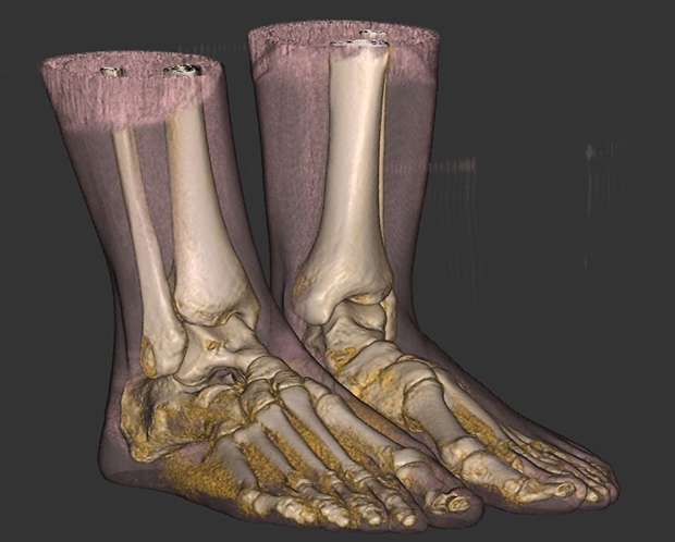
The pedCAT™, TRUE 3D weight-bearing imaging device was introduced during
2012 and recognized as one of the top ten innovations by Podiatry
Today. Unlike traditional CT scanners, the pedCAT™ allows the patient to
stand, making one revolution around the patient to capture the entire
region of interest.
This technology, known as cone beam volumetric tomography (CBVT), will
aid doctors in diagnosing and treating conditions including but not
limited to fractures, subluxations (misalignment) and dislocations,
midfoot injuries, bunions, flat feet, sprains, arthritis, and
diabetic-related complications. This CT is ideal for pre-planning,
post-operative planning, diagnosis of fractures, and evaluation of
arthritic joints, bunion deformities, ankle instability, foot alignment,
and sesamoid position and condition. Physicians benefit from this
technology because it provides a full view of the foot and ankle and
interactions of the bones, ligaments, and joints.
The Foot & Ankle Center of Illinois is the first practice in the
region to offer this new advanced diagnostic imaging to patients.
According to Dr. John Sigle, founder of the Foot & Ankle Center of
Illinois, “Having an in-office, state-of-the-art imaging system has
allowed us to reinvent our approach to surgical planning. Scans are
taken in a matter of minutes.
Scans provide a more targeted diagnosis. We use these three-dimensional
replicas to pre-plan implant surgeries, assuring a higher rate of
accuracy when screws, plates, and replacement joints are placed inside
the foot. We can also use scans to better assess arthritic joints and
detect bone erosion caused by diabetes. Advanced imaging enables us to
provide the right diagnosis at the right time resulting in better health
outcomes and lower costs.”
According to Dr. Grant Gonzalez, DPM, “The pedCAT™, TRUE 3D
weight-bearing imaging device is an advanced computer imaging system
that aids in complicated deformity correction. We are able to more
precisely plan and perform hindfoot reconstructions, flatfoot, and
multi-planar corrections with more accuracy. Pre-planning permits us to
reduce surgery time as we are able to execute a surgical plan more
effectively; we can simplify the most challenging portions of a
difficult case, making the process more efficient and more predictable.”
Dr. Sigle also uses this computer-guided surgical planning system on all
total ankle replacements, bunions, and hallux rigidus (stiff big toe)
procedures.
“We always have the best interest of our patients in mind with regards
to radiation exposure,” said Dr. Gonzalez. “One of the reasons we chose
the pedCAT™ was based on minimal radiation levels that are significantly
lower than traditional CT scanners.”
According to the Ludlow, J. International Journal of Diagnostic Imaging,
2014, a standing CT exposes you to about two to six micro Sieverts of
radiation. To put this in perspective, the average American is exposed
to about eight micro Sieverts of radiation a day from his or her
environment. A passenger on a flight from Los Angeles to New York is
exposed to about 40 micro Sieverts of radiation. Peer-reviewed studies
state the radiation dose of a standing CT is insignificant and should
not be a deciding factor when determining if a patient needs a scan.
New imaging CT scanners are being used to address complicated
conditions to provide more precise images and data for diagnosis and
surgery. Being able to measure and evaluate foot deformities in three
dimensions rather than two transforms diagnosis and drives better
surgical outcomes. If you are seeking consultation for foot or ankle
deformity, contact the Foot & Ankle Center of Illinois at
217-787-2700. The Foot & Ankle Center of Illinois is conveniently
located in Springfield, Decatur, Taylorville, Shelbyville, Sullivan, and
Carlinville. Visit myfootandanklecenter.com to view a short video of The Curve Beam pedCAT™, TRUE 3D weight bearing scanner.
There are numerous options to end heel pain. If you are seeking more
information or would like to schedule a consultation to end your heel
pain, call the Foot & Ankle Center of Illinois at 217-787-2700. We
have clinics located in Springfield, Taylorville, Decatur, Carlinville,
Shelbyville, and Sullivan. Visit myfootandanklecenter.com to obtain
information for stretching exercises you can do at home. Also, view a
short video on Cutting Edge MLS Laser Therapy and listen to physician
and patient testimonials about this new technology.