By Jeni Nenne, RT (R)(CV), OSF HealthCare St. Joseph Medical Center
A recent U.S. study reveals that four in 10 cases of breast cancer in younger women can be blamed on high breast density. The results show that breast density is a much more important breast cancer risk factor to be aware of than a person’s family history.
Having dense breast tissue is not an abnormal condition. In the United States, 46 percent of women have dense breast tissue. It’s basically a physical attribute of the body, and there’s little anyone can do to actively change or improve the density of their breast.
Breast tissue consists of fatty and fibroglandular tissue. Dense breast tissue is defined as having a higher percentage of fibroglandular tissue within your breasts. If more than 50 percent of your breast is made of fibroglandular tissue, then your breasts are classified as “dense.”
The more fibrous and glandular tissues absorb more radiation during mammography, reducing the accuracy of the test, making it more difficult to properly diagnose breast cancer. Dense breast tissue shows up white on a mammogram, as do tumors, which makes it more difficult to identify the differencae.
According to the American Congress of Obstetricians and Gynecologists, a mammogram will identify 88 percent of cancers in a breast that is almost entirely fat, or low in density. That’s compared to 62 percent in high-density breasts.
That’s where Automated Breast Ultrasound (ABUS) screening can provide a better evaluation for women with dense breasts. When used in addition to mammography, it can improve breast cancer detection by 55 percent over mammography alone.
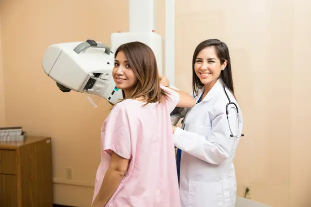
ABUS breast imaging’s 3D ultrasound technology uses sound waves instead of radiation. The sound waves create a 3D picture of the breast tissue. A radiologist can then review those pictures along with mammogram images. ABUS complements mammography but does not replace it. They work in different ways, and each has it owns attributes.
Women with dense breasts will have a mammogram that is very “white,” or difficult to see through. It is like looking through a snowstorm for a snowball — the tissues can all look the same. A cancer can easily hide in a background of dense breast tissue since they both appear white. Alternatively, in fatty tissue, which is gray, a white mass (cancer) can be readily identified. ABUS can help distinguish the dense tissue where a mass or lump appears as a dark area on a white background. With an order from a primary care provider, ABUS can be scheduled to be completed during the same appointment as a woman’s annual mammogram.
Women should talk with their health care provider about other breast cancer risk factors, such as family history. There are also lifestyle changes to consider. Reducing alcohol intake is one change as women who have more than one drink a day have an increased risk of breast cancer. Being overweight or obese after menopause also increases the risk, as fatty tissue makes estrogen. Even a small amount of weight loss can help reduce this risk.
Jeni Nenne, RT (R)(CV), is a Radiant Credentialed Trainer and Supervisor of Clinical Radiology Operations at OSF HealthCare St. Joseph Medical Center in Bloomington. To see if you would benefit from ABUS, call OSF St. Joseph College Avenue Imaging in Bloomington at 309-664-3272.